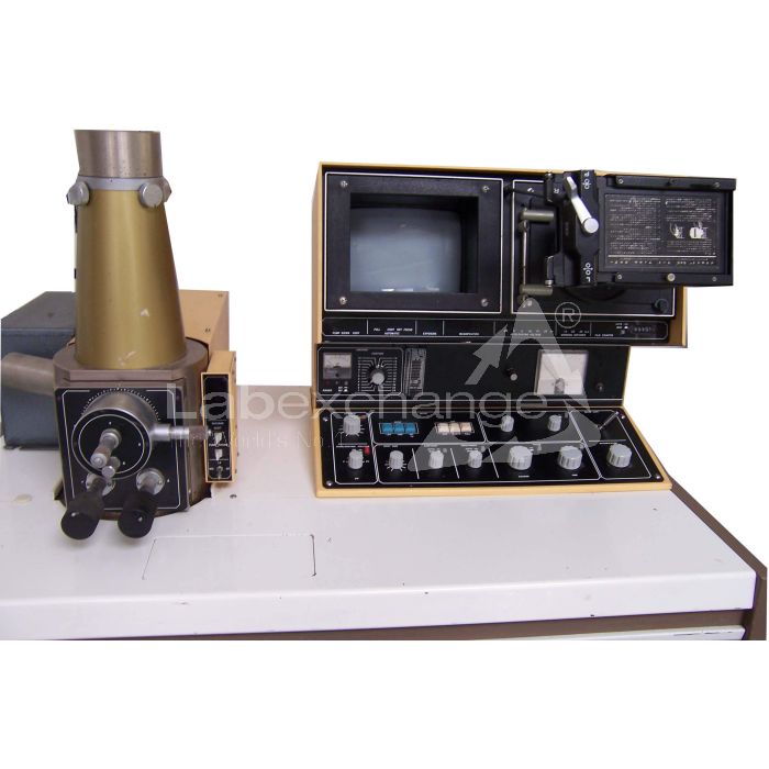
Jeol JSM T200
| Objektnummer | B00013303 |
|---|---|
| ID-Nummer | 013303 |
| Objektbezeichnung | Jeol JSM T200 |
| Status | Archiviertes Produkt |
Produktgruppe: Elektronenmikroskope
Status, Liefer- und Zahlungsbedingungen
Geräteüberprüfung
Die gebrauchten Laborgräte werden vor der Auslieferung von der Labexchange Service GmbH überprüft. Sie erhalten voll funktionsfähige Geräte.
Versandzeit
Die angegebenen Versandzeiten sind die jeweils kürzesten für einen Artikel. Die tatsächlich Versandzeiten können im Einzelfall davon abweichen. Die endgültigen Versandzeiten werden in der Auftragsbestätigung angegeben.
Bei Bestellung/Anfrage von mehreren Artikeln bieten wir grundsätzlich Sammellieferung an. Die Versandzeit berechnet sich nach der Position mit der längsten Versandzeit. Auf ausdrücklichen Wunsch ist eine Teillieferung möglich.
Versandarten
Paketdienste, Speditionen, Selbstabholung, Lieferung durch Labexchange-Fuhrpark
Lieferinformationen
Unsere Lieferbedingungen sind grundsätzlich zzgl. Versandkosten. Angegebene Versandkosten sind zu erwarten. Falls anfallende Versandkosten nicht angegeben sind, fragen Sie diese bitte gesondert an.
Die angegebenen Fracht- und Verpackungskosten beziehen sich auf den günstigsten Transportweg und sind vorbehaltlich unvorhergesehener Kostensteigerungen. Durch unvorhersehbare Ereignisse können sich die Frachtraten und die Lieferzeiten jederzeit ändern und müssten der aktuellen Situation angepasst werden. Incoterm-Kodierung gemäß Incoterms 2010: Bei Selbstabholung EXW, bei Sendungen per Schiff CFR, per Luftfracht CPT, übrige Sendungen DAP. Hinweis für Auslandssendungen: Ein Präferenznachweis/EUR1 wird von uns nicht ausgestellt. Bei Selbstabholung/EXW aus Drittländern und der EU werden 16% MWSt als Kaution einbehalten bis wir die Gelangensbestätigung/den Verbringungsnachweis des Käufers erhalten haben.
Zahlungsbedingungen
Wir akzeptieren keine Zahlung per Letter of Credit, PayPal etc. Der Rechnungsbetrag ist in jedem Fall ohne Abzug fällig. Die Ware bleibt bis zur vollständigen Bezahlung unser Eigentum. Skonto wird nicht gewährt.
|
Land |
Mögliche Zahlungsarten |
Bemerkung |
|
DE, AT, CH |
Rechnung, Vorkasse, Kreditkarte |
Eine Zahlung per Rechnung ist nur für Firmenkunden möglich. |
|
NL, BE, LU |
Rechnung, Vorkasse, Kreditkarte |
Eine Zahlung per Rechnung ist nur für Firmenkunden möglich. |
|
Alle weiteren Länder |
Vorkasse, Kreditkarte |
|
Es gelten unsere Allgemeinen Verkaufs-, Lieferungs- und Zahlungsbedingungen. Diese finden Sie hier. Zwischenverkauf, sowie Irrtum und Preisänderungen sind vorbehalten.
Statusdefinition
Alle Artikel sind gebrauchte Artikel, es sei denn ein Artikel wird explizit als Neugerät aufgeführt.
|
Status |
Zustand |
Bemerkung |
|
Sofort verfügbar |
gebraucht |
Der Artikel wurde bereits überprüft und befindet sich in einem einwandfreien Zustand. Er kann direkt an Sie versendet werden. |
| Lagergerät |
gebraucht |
Der Artikel befindet sich in unserem Lager. Unsere Techniker werden den Artikel vor der Auslieferung überprüfen. Sie erhalten voll funktionsfähige Artikel. |
|
Anbieter |
gebraucht |
Der Artikel befindet sich noch beim Anbieter. Nach Ihrer Bestellung wird er von uns angekauft, überprüft und an Sie versendet. Ein Funktionszertifikat und ein Servicebericht sind bei der Lieferung enthalten. |
|
Neugerät |
neu |
Es handelt sich um einen fabrikneuen Artikel. Es gelten die Garantiebestimmungen des Herstellers sowie die gesetzliche Gewährleistungsfrist. |
|
Labprocure |
gebraucht |
Verantwortlich für den Inhalt dieses Geräteangebotes ist die Labprocure GmbH als Geräteinserent. Labprocure übernimmt die Haftung für die hier inserierten Angebote und für die beinhalteten Fotos und Angebotstexte. Labprocure GmbH, Bruckstrasse 58, 72393 Burladingen. |
The following illustrations and descriptions are referring to the instrument model and are drawn from brochures. They are not representing the delivery volume. The exact delivery content you will find only in the offering text.
The Model T200 Scanning Microscope has been developed under the design philosophy of combining simple Operation, simple maintenance, and high performance. Accordingly, quality micrographs comparable to those obtainable by a large instrument can be readily obtained without any special skill.
These features together with the various advantages peculiar to the scanning microscope, such as very large depth of focus, wide magnification range, and minimal specimen preparation, make the T200 a most effective instrument for research work, quality control, and as a visual education aid.
GENERAL
The scanning electron microscope is a comparatively recent addition to the microscope family and is proving to be very popular along with the long established light and transmission type electron microscopes.
In spite of their merits and demerits, each type of microscope has its role to play depending on the field of application. For example, if a high resolving power and large depth of focus is not called for, a light microscope would be ideal. Where a very high resolving power is required, a transmission electron microscope would be necessary. However, when using a transmission electron microscope, the specimen must be very thin (less than 1 um (10,000 Ä) in thickness), a factor which requires a great deal of skill on the part of the user in order to prepare such specimens.
The scanning electron microscope, on the other hand, offers a fairly high resolution and moreover, since it is possible to use bulk specimens (specimen thickness being of no consequence), specimen preparation is easy. In addition to which, the depth of focus is large, thereby enabling 3-D observation.
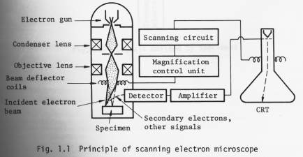
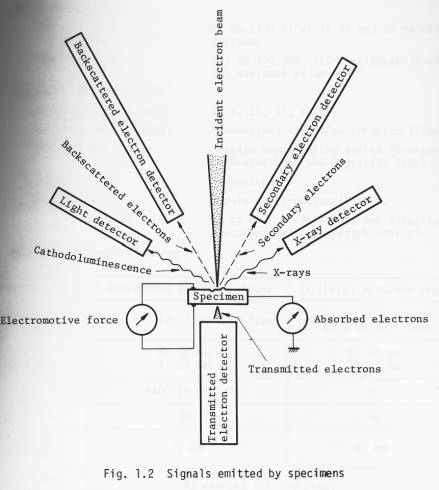
The operational principle of the scanning electron microscope is illustrated in Fig. 1.1. A finely focused electron probe is made to scan the specimen, resultant upon which, secondary and backscattered electrons, etc. (Fig. 1.2) are emitted from the surface of the specimen. These signals are then detected by a detector and outputted via an amplifier to a synchronously scanned CRT as an intensity variation signal. The CRT raster width divided by the electron probe scanning width gives the Image magnification.
By adding an appropriate detector (optional) to the Standard T200, transmitted electron images, cathodoluminescence images, etc. can be observed in addition to secondary and backscattered electron images. Further, by incorporating an X-ray detector, X-ray analysis becomes possible.
Specifications
Technical data
n Performance
Resolution: 10 nm (100 Ä) at 25 kV and 20 mm working distance.
Magnification: 15x to 100,000x (15x available at working distance 48 mm only).
n Electron optical system
Accelerating voltage: 2, 5, 10, 15, 25 kV.
Electron gun filament: Precentered cartridge tungsten filament.
Lens system: 3-stage demagnifying system (2-stage condenser lens and objective lens).
Alignment: Mechanical.
Stigmator: 8-pole electromagnetic type.
Image fine shift: Up to ±10 um (25 kV) in any direction; electromagnetic, joystick control.
n Specimen stage (twin stage)
|
Specimen stage |
I (Eucentric specimen stage) |
II (Large specimen stage) |
|
Specimen accomodation |
Up to 10 dia. x 10 thick*(mm) |
Up to 76.5dia. x 25.5 thick ** (mm) |
|
Range of movement |
X: 10 mm Y: 20 mm |
X: 40 mm Y: 40 mm |
|
Tilt |
-40° to +90°*** |
|
|
Rotation |
360° |
|
|
Working distance |
20 mm |
48 mm |
|
Specimen exchange |
By drawing out the stage |
|
|
Signal terminal |
Optional (max. 48 pins) |
|
* 32 dia., 51 dia., 76.5 dia. (mm) optionally available.
** Up to 127.5 dia. x 25.5 thick (mm) possible.
*** 220° possible.
n Scanning system
Secondary and backscattered
electron detection*: By a detector (comprizing a scintillator, a light pipe, a photomultiplier and a collector).
* Backscattered electron detector, which is capable of obtaining topographic and composition images, transmitted electron detector, cathodoluminescence detector, specimen current detector, X-ray
detector: Optionally available.
Scanning modes: Frame scan (including TV scan), line scan and Y modulation.
Scanning speeds: Visual TV scan; 0.2, 0.33, 10 sec/ frame.
Line scan 0.2, 0.33, 10 sec/frame.
Record 60 sec/frame.
Magnification: 35x to 100,000x (23 steps; series of 35,
50, 75, 100, 150, 200, 350, ).
(15x available at WD = 48 mm).
Viewing area: 135 mm x 180 mm.
Cathode ray tube: 230 mm, green phosphor CRT (used for viewing and recording*).
* A CRT exclusively used for recording is optionally available.
n TV signal output terminal : BNC-R connector; VTR available; composite video signal output; positive polarity; output voltage 1 Vp-p; use of 75 ohm coaxial cable; scanning
frequency horizontal: 15.75 kHz, vertical: 60 Hz.
n Recording System
(complete with electro-
magnetic shutter and
shutter button)
CSI-1: Standard; Brownie roll film; 1 to 1/2 photographing ratio.
CSI-2: Polaroid pack film; 1 to 3/4 photographing ratio (optional).
CSI-3: 35 mm roll film; 1 to 1/4 photographing ratio (optional).
CSI-4: Polaroid sheet film; 1 to l photographing ratio (optional).
n Vacuum system
Operation: Fully automatic.
Ultimate pressure: 7 x 10 -4 Pa (5 x 10 -6 Torr).
Vacuum gauge: Pirani gauge.
Pump-down time: About 30 minutes (from cold).
Specimen exchange: About 2.5 minutes.
Oil rotary pump: 100 l/min 1 unit.
Oil diffusion pump: 420 l/sec 1 unit.
n Safety devices: Devices for power failure, water failure and vacuum deterioration: built-in.
n Miscellaneous: Service outlet: built-in (100 V, 2 A; used for operating optional attachments).
The instrument can be wheeled an its own casters.
2. Installation requirements
n Power and water
Power: 100 V, 50/60 Hz, single phase, 2 kVA (basic instrument: 1.2 kVA; attachments: 0.8 kVA).
Starting current 60 A (0.2 sec.)
Fluctuation: less than ±10% (that also includes initial Operation).
Ground terminal: 1 terminal, less than 100 Ω.
Cooling water: Flow rate 2 l/min at 0.05 0.2 MPa (0.5 2 kg/cm 2 ).
Temperature 20 ±5 °C (water temperature at the outlet: not greater than 35 °C).
Faucet 1, 12 mm O.D.
Drain 1.
n Installation room
Room temperature: 20 ±5 °C.
Relative humidity: Less than 80%.
Floor vibration: Less than 2 µm p-p (5 Hz) in the X, Y and Z directions, less than 3 µm p-p (10 Hz) in the X, Y and Z directions, and less than 8 µm p-p (50 Hz) in the X, Y and Z directions (with the instrument installed).
Stray magnetic fields: Less than 0.3 µT (3 mG).
Note: The above specifications are subject to change without notice.
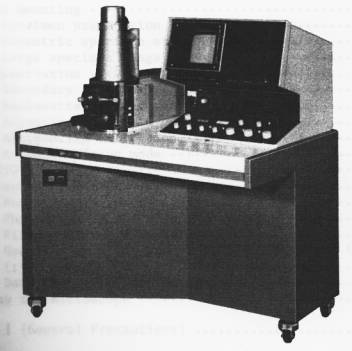
Layout, dimmensions and weight
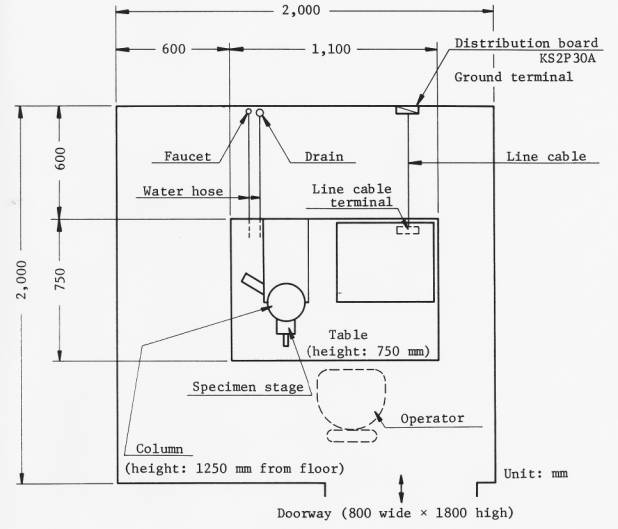
Overall weight (basic instrument: about 280 kg)
Fragen?
Ihr Labexchange-Team hilft gerne weiter:

Christian Schmid
Labor und Analytik, Laboreinrichtung, Life Science

Hubert Sauter
Spektroskopie, Chromatographie


