
Zeiss Axio Imager M1
| Objektnummer | B00018355 |
|---|---|
| ID-Nummer | 018355 |
| Objektbezeichnung | Zeiss Axio Imager M1 |
| Status | Archiviertes Produkt |
Produktgruppe: Mikroskope (Allgemein)
Status, Liefer- und Zahlungsbedingungen
Geräteüberprüfung
Die gebrauchten Laborgräte werden vor der Auslieferung von der Labexchange Service GmbH überprüft. Sie erhalten voll funktionsfähige Geräte.
Versandzeit
Die angegebenen Versandzeiten sind die jeweils kürzesten für einen Artikel. Die tatsächlich Versandzeiten können im Einzelfall davon abweichen. Die endgültigen Versandzeiten werden in der Auftragsbestätigung angegeben.
Bei Bestellung/Anfrage von mehreren Artikeln bieten wir grundsätzlich Sammellieferung an. Die Versandzeit berechnet sich nach der Position mit der längsten Versandzeit. Auf ausdrücklichen Wunsch ist eine Teillieferung möglich.
Versandarten
Paketdienste, Speditionen, Selbstabholung, Lieferung durch Labexchange-Fuhrpark
Lieferinformationen
Unsere Lieferbedingungen sind grundsätzlich zzgl. Versandkosten. Angegebene Versandkosten sind zu erwarten. Falls anfallende Versandkosten nicht angegeben sind, fragen Sie diese bitte gesondert an.
Die angegebenen Fracht- und Verpackungskosten beziehen sich auf den günstigsten Transportweg und sind vorbehaltlich unvorhergesehener Kostensteigerungen. Durch unvorhersehbare Ereignisse können sich die Frachtraten und die Lieferzeiten jederzeit ändern und müssten der aktuellen Situation angepasst werden. Incoterm-Kodierung gemäß Incoterms 2010: Bei Selbstabholung EXW, bei Sendungen per Schiff CFR, per Luftfracht CPT, übrige Sendungen DAP. Hinweis für Auslandssendungen: Ein Präferenznachweis/EUR1 wird von uns nicht ausgestellt. Bei Selbstabholung/EXW aus Drittländern und der EU werden 16% MWSt als Kaution einbehalten bis wir die Gelangensbestätigung/den Verbringungsnachweis des Käufers erhalten haben.
Zahlungsbedingungen
Wir akzeptieren keine Zahlung per Letter of Credit, PayPal etc. Der Rechnungsbetrag ist in jedem Fall ohne Abzug fällig. Die Ware bleibt bis zur vollständigen Bezahlung unser Eigentum. Skonto wird nicht gewährt.
|
Land |
Mögliche Zahlungsarten |
Bemerkung |
|
DE, AT, CH |
Rechnung, Vorkasse, Kreditkarte |
Eine Zahlung per Rechnung ist nur für Firmenkunden möglich. |
|
NL, BE, LU |
Rechnung, Vorkasse, Kreditkarte |
Eine Zahlung per Rechnung ist nur für Firmenkunden möglich. |
|
Alle weiteren Länder |
Vorkasse, Kreditkarte |
|
Es gelten unsere Allgemeinen Verkaufs-, Lieferungs- und Zahlungsbedingungen. Diese finden Sie hier. Zwischenverkauf, sowie Irrtum und Preisänderungen sind vorbehalten.
Statusdefinition
Alle Artikel sind gebrauchte Artikel, es sei denn ein Artikel wird explizit als Neugerät aufgeführt.
|
Status |
Zustand |
Bemerkung |
|
Sofort verfügbar |
gebraucht |
Der Artikel wurde bereits überprüft und befindet sich in einem einwandfreien Zustand. Er kann direkt an Sie versendet werden. |
| Lagergerät |
gebraucht |
Der Artikel befindet sich in unserem Lager. Unsere Techniker werden den Artikel vor der Auslieferung überprüfen. Sie erhalten voll funktionsfähige Artikel. |
|
Anbieter |
gebraucht |
Der Artikel befindet sich noch beim Anbieter. Nach Ihrer Bestellung wird er von uns angekauft, überprüft und an Sie versendet. Ein Funktionszertifikat und ein Servicebericht sind bei der Lieferung enthalten. |
|
Neugerät |
neu |
Es handelt sich um einen fabrikneuen Artikel. Es gelten die Garantiebestimmungen des Herstellers sowie die gesetzliche Gewährleistungsfrist. |
|
Labprocure |
gebraucht |
Verantwortlich für den Inhalt dieses Geräteangebotes ist die Labprocure GmbH als Geräteinserent. Labprocure übernimmt die Haftung für die hier inserierten Angebote und für die beinhalteten Fotos und Angebotstexte. Labprocure GmbH, Bruckstrasse 58, 72393 Burladingen. |
manufacturer: Zeiss
model: Axio Imager M1
annotation: Dokumente engl.
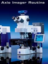
Axio Imager:
The New Versatility in Routine Microscopy
Efficiency, ergonomics, impressive application versatility and the best possible optics with these performance features, Axio Imager from Carl Zeiss is raising the standard in routine microscopy to a new level. Designed for research, Axio Imager combines the requirements and high performance of a high-end research microscope with easy handling and maximum convenience:
Outstanding optics with high resolution and excellent contrast
Well thought-out ergonomics concept and great ease of operation
Expandable modular system architecture manual or motorized
Specially designed stages for any application
For all optical techniques in transmitted and reflected light
Intuitive user guidance for image processing and documentation
The Ergonomics: The Concept for Relaxed Microscopy
Workplace microscope: this means, you have to spend long hours day after day on repetitive tasks at the microscope, putting an immense strain on both your body and your ability to concentrate. You really need the technology you use to be ergonomic. Carl Zeiss has designed an ergonomics concept for Axio Imager that relieves the strain as much as possible for the user: with convenient technology for fatigue-free, user-friendly operation. Allowing better concentration. Thus enabling you to be relaxed and more efficient in your work.
The consequences of a poor viewing height
Ergonomic studies prove that the conventional microscope height and, therefore, the height for looking into the eyepiece is too low for the overwhelming majority of users involved in routine microscopy. The user has to adjust the position of his body to suit this low viewing height. This brings with it a whole host of consequences that all put a major strain on the body. The forearms rest on the edge of the table, causing painful pressure points and making it difficult to operate the controls. The head and back are bent forward. Muscles tense up under the immense strain. Merely adjusting the viewing height using a swiveling eyepiece does not greatly improve the situation, as this puts the neck in a bad position in the upper height range. In short, to be able to work ergonomically with a table microscope, you need to be sitting correctly, with your body virtually straight, and looking into the eyepiece at the right angle.
Convenient viewing: the ergotube
The crucial element here is individual adjustment thanks to a height-adjustable tube. Axio Imager requires minimal tilting of the head when the user is looking into the eyepiece, taking the strain off the neck and shoulder muscles. It achieves this with the help of the new ergotube: with 50 mm height adjustment, a fixed viewing angle of 15° (which is ergonomically ideal) and an eyepiece extension of 50 mm, it can be individually adjusted to suit the size of any user. The body remains completely relaxed, even during long microscopy sessions.
Fields of Application: Boundless Possibilities for Routine Microscopy
Axio Imager offers the user a system platform that supports all requirements, from brightfield applications and polarization to complex FISH applications. And with a modular architecture that is tailored to growing demands. Application-specific components are the perfect addition to the basic features of all Axio Imager stand variants. A sound investment for now and for the future.
Pathology
Axio Imager.A1 with LED illumination is ideal for pathology. It features a manual stand with Fixed-Koehler illumination. Used in conjunction with Achroplan or EC Plan-Neofluar objectives, this is the basic equipment required for histological evaluation. The cost-effective LED illumination is durable, uses little energy and requires no maintenance or adjustment. It produces impressive images when used with the typical H.-E., DAB or Azan stains. Its constant color temperature ensures consistent light quality and a brilliant image across the entire adjustment range. The specially designed stage for pathology with its white
Pathology with polarization contrast
Polarization contrast is essential for detecting foreign bodies in tissue or for diagnosing Alzheimers, for example, and is therefore a basic feature of any pathology microscope. Depending on the application, fixed or rotatable polarizers and analyzers can be used, or even a wave plate. Axio Imager.A1 with LED illumination is perfectly equipped for simple tasks. Particularly suitable for this application are the EC Plan-Neofluar and Plan-Apochromat objectives from Carl Zeiss.
Histology and anatomy
The requirements in histology and anatomy include optimum resolution in the image viewed and digitally captured, perfect color display in the documentation of details and overviews, and fast, precise relocalization of meaningful positions on the specimen. Ideal for this purpose are the EC Plan-Neofluar and Plan-Apochromat objectives combined with motorized stages.
Immunocytochemistry with reflection contrast
The evaluation of immunocytochemically stained specimens not only calls for the stability and specificity of the labeling; an excellent color display and high resolution are also extremely important. For this reason, people involved in routine microscopy like to use immunogold methods (IGS or IGSS), which are analyzed in reflection contrast. This reflected light contrast is produced with polarized light and can be displayed ideally using the EC Plan-Neofluar 63x/1.25 Oil Ph3 Antiflex objective. A quarter-wave plate integrated into the objective provides an optimum setting, for brilliant contrast, perfect homogeneity and unprecedented resolution. Reflection contrast also makes it possible to display distances and points of adherence between cells and their substrate in the nm range, enabling immunocytochemical labeling methods to be coupled with cell adhesion assays.
Diagnostic cytology
Diagnostic cytology is a routine procedure in e.g., gynecology and tumor diagnostics. Typical stain: Papanicolaou. Using the objectives in the A-Plan category, the Achroplan 50x/0.9 for covered and uncovered specimens as well as the Plan-Apochromat 63x/1.4 Oil, and Axio Imager, you can achieve a diagnosis even more quickly. Depending on the application, you can add a mechanical stage with an object marker, a pathologists stage, a stage with electronic verniers, or a motorized stage.
Hygiene (Parasitology, Mikrobiology, Virology)
Axio Imager is the perfect tool for detecting parasites, bacteria or virus aggregates with the high resolution and enhanced-contrast EC Plan-Neofluar objectives. The motorized Axio Imager.M1 offers particular benefits for routine microscopy, with its motorized switching between the different transmitted light contrast methods and reflected-light fluorescence using the contrast and light managers. The illumination intensity and contrast are always adjusted optimally for the chosen magnification. Typical uses: diagnosing malaria or borreliosis.
Genetics with FISH applications
In genetics, genome analyses (karyograms) are often documented and analyzed in transmitted light brightfield to diagnose illnesses that are attributable to a genotype mutation. The Fluorescence-In-Situ-Hybridization (FISH) method identifies the gene loci on the chromosomes with the help of the DNA probes used and helps to track deviations from a healthy state very quickly. Axio Imager offers full support in this process: the apochromatically-corrected IC 2 S beam path illuminates the object field evenly for all colors. The integrated light-traps purposefully eliminate stray light in the illumination and imaging beam paths.
The 6x reflector turrets for Axio Imager.A1 and M1, as well as the 10x reflector turrets for Axio Imager.D1 and Z1, enable rapid multichannel image acquisition the basis for FISH analyses. AxioVision or MetaCyte (by Metasystems) control makes handling such complex applications as simple as possible.
Telepathology with Axio Imager: four eyes see more than two
Whether we are talking about quick section evaluation, obtaining a second opinion or consultation within a network of experts online specimen evaluation is an extension to traditional microscopy in all areas of application. Where time or cost is a key factor in evaluating specimens, or the pathologist or his co-workers cannot perform an evaluation on site, it has to be possible to carry out an analysis live and by remote operation.
Axio Imager the ideal platform
The performance features of Axio Imager are perfect for telepathology: an intelligent motorization and operating concept, maximum optical efficiency, the possibility of expansion and a large range of uses. These parameters make Axio Imager the most efficient microscope system for telepathology. The software satisfies all requirements, from data transmission to data security. Used in combination with AxioVision, Axio Imager offers high-resolution digital images for diagnosing on the system.
Within easy reach: the control buttons
Operating the system is as ergonomic as it is convenient: all the control buttons are arranged around the focus drive. Tactile surfaces make it easy to tell them apart. On the manual stand, you can use five preconfigured buttons to adjust light intensity and operate the motorized shutters in reflected and transmitted light. And for those who want more convenience, there are ten freely configurable buttons available on the motorized stand.
All the functions at a glance: the touch screen
On the motorized stand, all the functions are grouped together on the touch screen (TFT display): you can control all motorized components and view the microscope status simply by pressing with your finger, making complex work steps significantly easier and quicker. If necessary, they can also be stored as settings and retrieved at any time at the touch of a button.
Documentation with AxioVision: Achieving Precise Results Faster
AxioVision from Carl Zeiss is the modular software platform for all digital documentation requirements in routine microscopy. From simple image acquisition to image analysis, AxioVision supports any application by offering a large number of functions that can be automated as well as clear user guidance. And if your requirements grow, AxioVision can also be expanded with numerous add-on modules.
Reproducibility due to automatic scaling
Once the objectives have been configured and scaled in Axio Imager, AxioVision automatically assigns the appropriate scaling. The automatic scaling reduces the statistical human measurement error to a minimum and is perfectly reproducible every time. The net result is that operation is much simpler and your scientific results are more reliable.
Crucial in routine microscopy: simple measurement
Even with the basic version of AxioVision, it is possible to define object-describing parameters interactively for simple measurements. Typical applications include measuring distances (e.g., the spread of focuses of inflammation) and areas (e.g., the size of cells), or counting cell nuclei in a particular area.
For large surfaces: MosaiX and Panorama
Large specimens can be acquired and analyzed very easily using the Panorama and MosaiX modules. The specimen is scanned in image field by image field on a manual or motorized mechanical/scanning stage. AxioVision generates a large composite image from the individual images. This then serves as a map for navigating around the sample, or as a basis for further analysis. If this joining of individual image fields based on coordinates is not sufficient, the Panorama module enables the individual component images to be combined to create a high-resolution Panorama image.
Fragen?
Ihr Labexchange-Team hilft gerne weiter:

Christian Schmid
Labor und Analytik, Laboreinrichtung, Life Science

Hubert Sauter
Spektroskopie, Chromatographie


