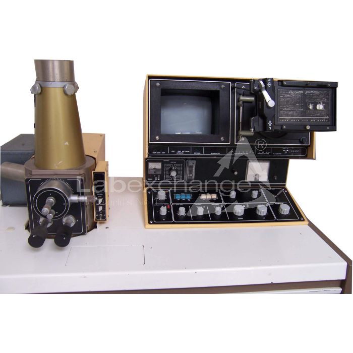
Jeol JSM T200
| Objektnummer | B00013303 |
|---|---|
| Seriennummer | 013303 |
| Object Naam | Jeol JSM T200 |
| Status | Gearchiveerd product |
Product groep: Electronenmicroscopen
Status, leverings- en betalingsvoorwaarden
Apparatuurcontrole
De gebruikte apparatuur wordt voorafgaand aan levering gecontroleerd door Labexchange Service GmbH. U ontvangt volledig functionerende apparatuur.
Verzending
De vermelde verzendtijden zijn telkens de kortste voor een artikel. In bepaalde gevallen kunnen de daadwerkelijke verzendtijden daarvan afwijken. De uiteindelijke verzendtijden worden aangegeven in de opdrachtbevestiging.
In de regel bieden we combinatieleveringen aan. Levertijden zijn afhankelijk van het artikel met de langste levertijd. Deelleveringen zijn mogelijk tegen een toeslag.
Verzendmethoden
Koeriersdiensten, transportbedrijven, zelf afhalen, levering door Labexchange wagenpark
Informatie levering
De prijzen zijn exclusief verzendkosten. De genoemde verzendkosten zijn de te verwachten kosten. Afwijkingen zijn mogelijk. In het geval geen kosten voor verzending zijn gespecificeerd, vraag die dan afzonderlijk aan.
De opgegeven vracht- en verpakkingskosten hebben betrekking op de goedkoopste transportroute en zijn onder voorbehoud van onvoorziene kostenstijgingen. Door onvoorziene gebeurtenissen kunnen vrachttarieven en levertijden op elk moment veranderen en moeten ze worden aangepast aan de huidige situatie. Incoterm coderingen volgens Incoterms 2010: Bij afhalen EXW, CFR voor zendingen over zee, CPT per luchtfracht, andere zendingen DAP. Opmerking: We geven geen preferentieel certificaat/EUR1 af. Bij zelf afhalen/af fabriek (EXW) uit derde landen en de EU wordt 16% btw als borg ingehouden, tot we de ontvangstbevestiging/het leveringscertificaat van de koper hebben ontvangen.
Betalingsvoorwaarden
Wij accepteren geen betalingen Letter of Credit, PayPal etc. Het factuurbedrag is volledig verschuldigd. Er zijn geen betalingskortingen. De goederen blijven tot volledige betaling ons eigendom.
|
Land |
Mogelijke betaalmethoden |
Opmerking |
|
Duitsland, Oostenrijk, Zwitserland |
Betaling via factuur, vooruitbetaling, per creditkaart |
Betaling via factuur is mogelijk voor ondernemingsklanten. |
|
Nederland, België en Luxemburg |
Betaling via factuur, vooruitbetaling, per creditkaart |
Betaling via factuur is mogelijk voor ondernemingsklanten. |
|
Andere landen |
vooruitbetaling, per creditkaart |
|
Onze Algemene Voorwaarden voor Verkoop, Levering en Betaling zijn hierop van toepassing. Deze voorwaarden zijn hier te downloaden.
Tussenverkoop is ons voorbehouden.
Beschrijving status:
Alle artikelen zijn gebruikte artikelen, tenzij bij een artikel uitdrukkelijk wordt vermeld dat het om een nieuw apparaat gaat.
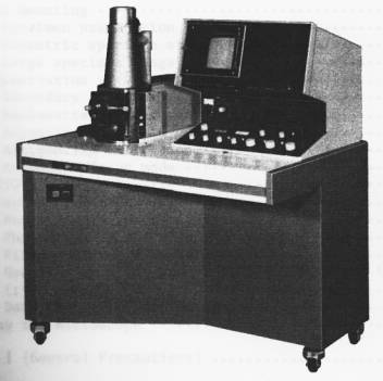
The following illustrations and descriptions are referring to the instrument model and are drawn from brochures. They are not representing the delivery volume. The exact delivery content you will find only in the offering text.
The Model T200 Scanning Microscope has been developed under the design philosophy of combining simple Operation, simple maintenance, and high performance. Accordingly, quality micrographs comparable to those obtainable by a large instrument can be readily obtained without any special skill.
These features together with the various advantages peculiar to the scanning microscope, such as very large depth of focus, wide magnification range, and minimal specimen preparation, make the T200 a most effective instrument for research work, quality control, and as a visual education aid.
GENERAL
The scanning electron microscope is a comparatively recent addition to the microscope family and is proving to be very popular along with the long established light and transmission type electron microscopes.
In spite of their merits and demerits, each type of microscope has its role to play depending on the field of application. For example, if a high resolving power and large depth of focus is not called for, a light microscope would be ideal. Where a very high resolving power is required, a transmission electron microscope would be necessary. However, when using a transmission electron microscope, the specimen must be very thin (less than 1 um (10,000 Ä) in thickness), a factor which requires a great deal of skill on the part of the user in order to prepare such specimens.
The scanning electron microscope, on the other hand, offers a fairly high resolution and moreover, since it is possible to use bulk specimens (specimen thickness being of no consequence), specimen preparation is easy. In addition to which, the depth of focus is large, thereby enabling 3-D observation.
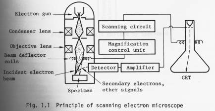
The operational principle of the scanning electron microscope is illustrated in Fig. 1.1. A finely focused electron probe is made to scan the specimen, resultant upon which, secondary and backscattered electrons, etc. (Fig. 1.2) are emitted from the surface of the specimen. These signals are then detected by a detector and outputted via an amplifier to a synchronously scanned CRT as an intensity variation signal. The CRT raster width divided by the electron probe scanning width gives the Image magnification.
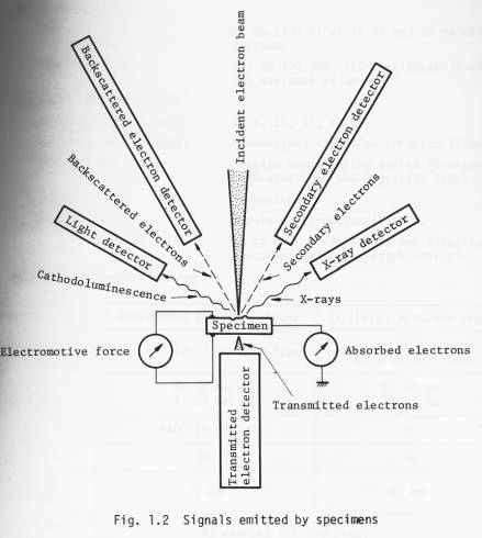
By adding an appropriate detector (optional) to the Standard T200, transmitted electron images, cathodoluminescence images, etc. can be observed in addition to secondary and backscattered electron images. Further, by incorporating an X-ray detector, X-ray analysis becomes possible.
Specifications
Technical data
n Performance
Resolution: 10 nm (100 Ä) at 25 kV and 20 mm working distance.
Magnification: 15x to 100,000x (15x available at working distance 48 mm only).
n Electron optical system
Accelerating voltage: 2, 5, 10, 15, 25 kV.
Electron gun filament: Precentered cartridge tungsten filament.
Lens system: 3-stage demagnifying system (2-stage condenser lens and objective lens).
Alignment: Mechanical.
Stigmator: 8-pole electromagnetic type.
Image fine shift: Up to ±10 um (25 kV) in any direction; electromagnetic, joystick control.
n Specimen stage (twin stage)
|
Specimen stage |
I (Eucentric specimen stage) |
II (Large specimen stage) |
|
Specimen accomodation |
Up to 10 dia. x 10 thick*(mm) |
Up to 76.5dia. x 25.5 thick ** (mm) |
|
Range of movement |
X: 10 mm Y: 20 mm |
X: 40 mm Y: 40 mm |
|
Tilt |
-40° to +90°*** |
|
|
Rotation |
360° |
|
|
Working distance |
20 mm |
48 mm |
|
Specimen exchange |
By drawing out the stage |
|
|
Signal terminal |
Optional (max. 48 pins) |
|
* 32 dia., 51 dia., 76.5 dia. (mm) optionally available.
** Up to 127.5 dia. x 25.5 thick (mm) possible.
*** 220° possible.
n Scanning system
Secondary and backscattered
electron detection*: By a detector (comprizing a scintillator, a light pipe, a photomultiplier and a collector).
* Backscattered electron detector, which is capable of obtaining topographic and composition images, transmitted electron detector, cathodoluminescence detector, specimen current detector, X-ray
detector: Optionally available.
Scanning modes: Frame scan (including TV scan), line scan and Y modulation.
Scanning speeds: Visual TV scan; 0.2, 0.33, 10 sec/ frame.
Line scan 0.2, 0.33, 10 sec/frame.
Record 60 sec/frame.
Magnification: 35x to 100,000x (23 steps; series of 35,
50, 75, 100, 150, 200, 350, ).
(15x available at WD = 48 mm).
Viewing area: 135 mm x 180 mm.
Cathode ray tube: 230 mm, green phosphor CRT (used for viewing and recording*).
* A CRT exclusively used for recording is optionally available.
n TV signal output terminal : BNC-R connector; VTR available; composite video signal output; positive polarity; output voltage 1 Vp-p; use of 75 ohm coaxial cable; scanning
frequency horizontal: 15.75 kHz, vertical: 60 Hz.
n Recording System
(complete with electro-
magnetic shutter and
shutter button)
CSI-1: Standard; Brownie roll film; 1 to 1/2 photographing ratio.
CSI-2: Polaroid pack film; 1 to 3/4 photographing ratio (optional).
CSI-3: 35 mm roll film; 1 to 1/4 photographing ratio (optional).
CSI-4: Polaroid sheet film; 1 to l photographing ratio (optional).
n Vacuum system
Operation: Fully automatic.
Ultimate pressure: 7 x 10 -4 Pa (5 x 10 -6 Torr).
Vacuum gauge: Pirani gauge.
Pump-down time: About 30 minutes (from cold).
Specimen exchange: About 2.5 minutes.
Oil rotary pump: 100 l/min 1 unit.
Oil diffusion pump: 420 l/sec 1 unit.
n Safety devices: Devices for power failure, water failure and vacuum deterioration: built-in.
n Miscellaneous: Service outlet: built-in (100 V, 2 A; used for operating optional attachments).
The instrument can be wheeled an its own casters.
2. Installation requirements
n Power and water
Power: 100 V, 50/60 Hz, single phase, 2 kVA (basic instrument: 1.2 kVA; attachments: 0.8 kVA).
Starting current 60 A (0.2 sec.)
Fluctuation: less than ±10% (that also includes initial Operation).
Ground terminal: 1 terminal, less than 100 Ω.
Cooling water: Flow rate 2 l/min at 0.05 0.2 MPa (0.5 2 kg/cm 2 ).
Temperature 20 ±5 °C (water temperature at the outlet: not greater than 35 °C).
Faucet 1, 12 mm O.D.
Drain 1.
n Installation room
Room temperature: 20 ±5 °C.
Relative humidity: Less than 80%.
Floor vibration: Less than 2 µm p-p (5 Hz) in the X, Y and Z directions, less than 3 µm p-p (10 Hz) in the X, Y and Z directions, and less than 8 µm p-p (50 Hz) in the X, Y and Z directions (with the instrument installed).
Stray magnetic fields: Less than 0.3 µT (3 mG).
Note: The above specifications are subject to change without notice.
Layout, dimmensions and weight
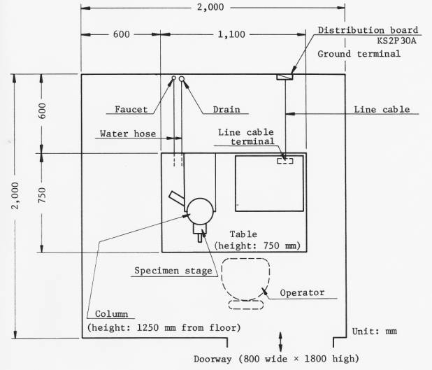
Overall weight (basic instrument: about 280 kg)
Onze Labexchange-team zullen u graag helpen:

Christian Schmid
Laboratorium en analytiek, Laboratoriuminrichting, Life science

Hubert Sauter
Spectroscopie, Chromatografie


