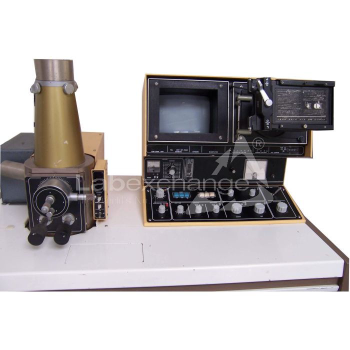
Jeol JSM T200
| Objektnummer | B00013303 |
|---|---|
| Seriennummer | 013303 |
| Nome oggetto | Jeol JSM T200 |
| Stato | Archived Product |
Gruppo prodotti: Microscopi Elettronici
Status, terms of delivery and payment
Verification of devices
The second-hand devices are verified by Labexchange Service GmbH before delivery. You are receiving only fully functional devices.
Dispatch time
The stated dispatch times are the shortest possible ones for each article. The effective dispatch times can vary. The effective dispatch times will be stated in the order confirmation.
As a matter of principle, we are offering collective deliveries. The shipping time is calculated based on the position with the longest lead time. A partial delivery is possible on explicit request.
Shipping methods
Parcel services, forwarding agencies, self-pickup, delivery by Labexchange fleet.
Delivery information
Prices exclude shipping costs. Stated shipping costs are to be expected. Deviations are possible. If transport costs are not specified, please ask separately for them.
The stated transport and packing charges apply to the most favorable route if transport and are to be understood as subject to verification due to unexpected cost increases. By reason of unpredictable events, cargo rates and delivery times can change at any time and therefore have to be adapted to the recent situation. Import formalities and possible customs charges will be borne by the purchaser. Incoterm coding according to Incoterms 2010: For persons who collect the devices themselves: EXW, for dipatch by sea: CFR, by air freight: CPT, other shipments: DAP. Note for international shipments: A proof of preference/EUR1 will not be issued by us. When self-collecting/ordering EXW from countries within or outside the European Union, 16% VAT will be retained as a deposit until we have received the corresponding confirmation of arrival/bill of delivery from the buyer.
Terms of payment
We do not accept payment by letter of credit, PayPal, etc. In each case the invoice amount is payable without deduction. Discount is not granted.
|
Country |
Possible payment methods |
Comment |
|
DE, AT, CH |
Payment by invoice, payment in advance, payment by credit card |
Payment by invoice is only possible for corporate clients. |
|
NL, BE, LU |
Payment by invoice, payment in advance, payment by credit card |
Payment by invoice is only possible for corporate clients |
|
Other countries |
Payment in advance, payment by credit card |
|
Our General Terms of Sale, Delivery and Payment are valid and are available for download here.
The goods are offered subject to prior sale.
Definition of status
All articles are used articles, except an article is listed especially as a new device.
|
Status |
Condition |
Comment |
|
Immediately available |
Used | The article is fully functional and in impeccable condition. It can be shipped immediately. |
| In stock |
Used |
The article is on stock. Our service technicians will verify the article before delivery. You are receiving only a fully functional article. |
|
Published |
Used |
The article is still with the provider. After your order the article will be purchased and verified by us before being shipped to you. A certificate of operativeness as well as a service report are included in delivery. |
|
New device |
new |
The article is brand new and unused. Regarding new equipment the guarantee/warranty conditions of the corresponding manufacturer apply. |
|
Labprocure |
Used |
Labprocure GmbH, as the advertiser, is responsible for the content of this device offer. Labprocure assumes liability for the offers advertised here and for the photos and offer texts included. Labprocure GmbH, Bruckstraße 58, 72393 Burladingen. |
The following illustrations and descriptions are referring to the instrument model and are drawn from brochures. They are not representing the delivery volume. The exact delivery content you will find only in the offering text.
The Model T200 Scanning Microscope has been developed under the design philosophy of combining simple Operation, simple maintenance, and high performance. Accordingly, quality micrographs comparable to those obtainable by a large instrument can be readily obtained without any special skill.
These features together with the various advantages peculiar to the scanning microscope, such as very large depth of focus, wide magnification range, and minimal specimen preparation, make the T200 a most effective instrument for research work, quality control, and as a visual education aid.
GENERAL
The scanning electron microscope is a comparatively recent addition to the microscope family and is proving to be very popular along with the long established light and transmission type electron microscopes.
In spite of their merits and demerits, each type of microscope has its role to play depending on the field of application. For example, if a high resolving power and large depth of focus is not called for, a light microscope would be ideal. Where a very high resolving power is required, a transmission electron microscope would be necessary. However, when using a transmission electron microscope, the specimen must be very thin (less than 1 um (10,000 Ä) in thickness), a factor which requires a great deal of skill on the part of the user in order to prepare such specimens.
The scanning electron microscope, on the other hand, offers a fairly high resolution and moreover, since it is possible to use bulk specimens (specimen thickness being of no consequence), specimen preparation is easy. In addition to which, the depth of focus is large, thereby enabling 3-D observation.
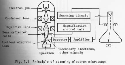
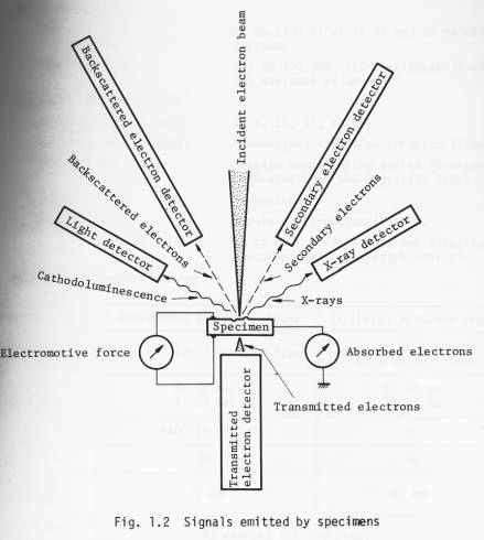
The operational principle of the scanning electron microscope is illustrated in Fig. 1.1. A finely focused electron probe is made to scan the specimen, resultant upon which, secondary and backscattered electrons, etc. (Fig. 1.2) are emitted from the surface of the specimen. These signals are then detected by a detector and outputted via an amplifier to a synchronously scanned CRT as an intensity variation signal. The CRT raster width divided by the electron probe scanning width gives the Image magnification.
By adding an appropriate detector (optional) to the Standard T200, transmitted electron images, cathodoluminescence images, etc. can be observed in addition to secondary and backscattered electron images. Further, by incorporating an X-ray detector, X-ray analysis becomes possible.
Specifications
Technical data
n Performance
Resolution: 10 nm (100 Ä) at 25 kV and 20 mm working distance.
Magnification: 15x to 100,000x (15x available at working distance 48 mm only).
n Electron optical system
Accelerating voltage: 2, 5, 10, 15, 25 kV.
Electron gun filament: Precentered cartridge tungsten filament.
Lens system: 3-stage demagnifying system (2-stage condenser lens and objective lens).
Alignment: Mechanical.
Stigmator: 8-pole electromagnetic type.
Image fine shift: Up to ±10 um (25 kV) in any direction; electromagnetic, joystick control.
n Specimen stage (twin stage)
|
Specimen stage |
I (Eucentric specimen stage) |
II (Large specimen stage) |
|
Specimen accomodation |
Up to 10 dia. x 10 thick*(mm) |
Up to 76.5dia. x 25.5 thick ** (mm) |
|
Range of movement |
X: 10 mm Y: 20 mm |
X: 40 mm Y: 40 mm |
|
Tilt |
-40° to +90°*** |
|
|
Rotation |
360° |
|
|
Working distance |
20 mm |
48 mm |
|
Specimen exchange |
By drawing out the stage |
|
|
Signal terminal |
Optional (max. 48 pins) |
|
* 32 dia., 51 dia., 76.5 dia. (mm) optionally available.
** Up to 127.5 dia. x 25.5 thick (mm) possible.
*** 220° possible.
n Scanning system
Secondary and backscattered
electron detection*: By a detector (comprizing a scintillator, a light pipe, a photomultiplier and a collector).
* Backscattered electron detector, which is capable of obtaining topographic and composition images, transmitted electron detector, cathodoluminescence detector, specimen current detector, X-ray
detector: Optionally available.
Scanning modes: Frame scan (including TV scan), line scan and Y modulation.
Scanning speeds: Visual TV scan; 0.2, 0.33, 10 sec/ frame.
Line scan 0.2, 0.33, 10 sec/frame.
Record 60 sec/frame.
Magnification: 35x to 100,000x (23 steps; series of 35,
50, 75, 100, 150, 200, 350, ).
(15x available at WD = 48 mm).
Viewing area: 135 mm x 180 mm.
Cathode ray tube: 230 mm, green phosphor CRT (used for viewing and recording*).
* A CRT exclusively used for recording is optionally available.
n TV signal output terminal : BNC-R connector; VTR available; composite video signal output; positive polarity; output voltage 1 Vp-p; use of 75 ohm coaxial cable; scanning
frequency horizontal: 15.75 kHz, vertical: 60 Hz.
n Recording System
(complete with electro-
magnetic shutter and
shutter button)
CSI-1: Standard; Brownie roll film; 1 to 1/2 photographing ratio.
CSI-2: Polaroid pack film; 1 to 3/4 photographing ratio (optional).
CSI-3: 35 mm roll film; 1 to 1/4 photographing ratio (optional).
CSI-4: Polaroid sheet film; 1 to l photographing ratio (optional).
n Vacuum system
Operation: Fully automatic.
Ultimate pressure: 7 x 10 -4 Pa (5 x 10 -6 Torr).
Vacuum gauge: Pirani gauge.
Pump-down time: About 30 minutes (from cold).
Specimen exchange: About 2.5 minutes.
Oil rotary pump: 100 l/min 1 unit.
Oil diffusion pump: 420 l/sec 1 unit.
n Safety devices: Devices for power failure, water failure and vacuum deterioration: built-in.
n Miscellaneous: Service outlet: built-in (100 V, 2 A; used for operating optional attachments).
The instrument can be wheeled an its own casters.
2. Installation requirements
n Power and water
Power: 100 V, 50/60 Hz, single phase, 2 kVA (basic instrument: 1.2 kVA; attachments: 0.8 kVA).
Starting current 60 A (0.2 sec.)
Fluctuation: less than ±10% (that also includes initial Operation).
Ground terminal: 1 terminal, less than 100 Ω.
Cooling water: Flow rate 2 l/min at 0.05 0.2 MPa (0.5 2 kg/cm 2 ).
Temperature 20 ±5 °C (water temperature at the outlet: not greater than 35 °C).
Faucet 1, 12 mm O.D.
Drain 1.
n Installation room
Room temperature: 20 ±5 °C.
Relative humidity: Less than 80%.
Floor vibration: Less than 2 µm p-p (5 Hz) in the X, Y and Z directions, less than 3 µm p-p (10 Hz) in the X, Y and Z directions, and less than 8 µm p-p (50 Hz) in the X, Y and Z directions (with the instrument installed).
Stray magnetic fields: Less than 0.3 µT (3 mG).
Note: The above specifications are subject to change without notice.
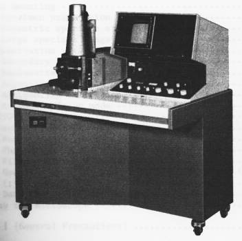
Layout, dimmensions and weight
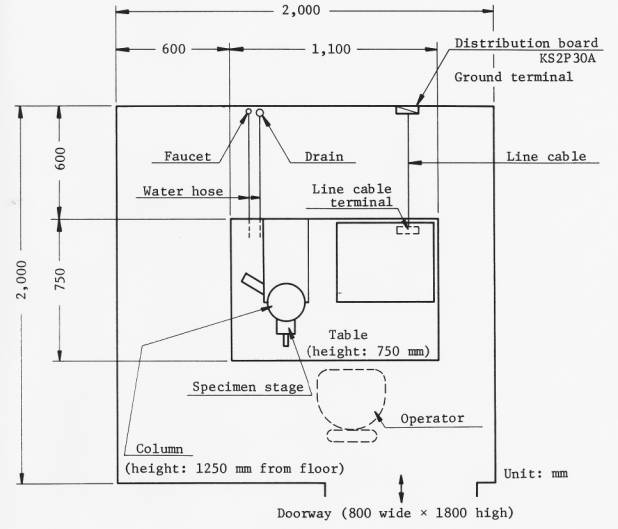
Overall weight (basic instrument: about 280 kg)
Il tuo Labexchange-Team sarà lieto di aiutarvi:

Christian Schmid
Laboratory and analysis, Laboratory equipment, Life science

Hubert Sauter
Spectroscopy, Chromatography


