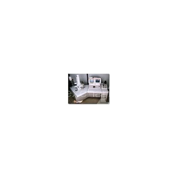
Jeol JSM-5400
Objektnummer | B00020299 |
---|---|
Seriennummer | 020299 |
Nome oggetto | Jeol JSM-5400 |
Stato | Archiv |
Status, terms of delivery and payment
Verification of devices
The second-hand devices are verified by Labexchange Service GmbH before delivery. You are receiving only fully functional devices.
Dispatch time
The stated dispatch times are the shortest possible ones for each article. The effective dispatch times can vary. The effective dispatch times will be stated in the order confirmation.
As a matter of principle, we are offering collective deliveries. The shipping time is calculated based on the position with the longest lead time. A partial delivery is possible on explicit request.
Shipping methods
Parcel services, forwarding agencies, self-pickup, delivery by Labexchange fleet.
Delivery information
Prices exclude shipping costs. Stated shipping costs are to be expected. Deviations are possible. If transport costs are not specified, please ask separately for them.
The stated transport and packing charges apply to the most favorable route if transport and are to be understood as subject to verification due to unexpected cost increases. By reason of unpredictable events, cargo rates and delivery times can change at any time and therefore have to be adapted to the recent situation. Import formalities and possible customs charges will be borne by the purchaser. Incoterm coding according to Incoterms 2010: For persons who collect the devices themselves: EXW, for dipatch by sea: CFR, by air freight: CPT, other shipments: DAP. Note for international shipments: A proof of preference/EUR1 will not be issued by us. When self-collecting/ordering EXW from countries within or outside the European Union, 16% VAT will be retained as a deposit until we have received the corresponding confirmation of arrival/bill of delivery from the buyer.
Terms of payment
We do not accept payment by letter of credit, PayPal, etc. In each case the invoice amount is payable without deduction. Discount is not granted.
Country |
Possible payment methods |
Comment |
DE, AT, CH |
Payment by invoice, payment in advance, payment by credit card |
Payment by invoice is only possible for corporate clients. |
NL, BE, LU |
Payment by invoice, payment in advance, payment by credit card |
Payment by invoice is only possible for corporate clients |
Other countries |
Payment in advance, payment by credit card |
|
Our General Terms of Sale, Delivery and Payment are valid and are available for download here.
The goods are offered subject to prior sale.
Definition of status
All articles are used articles, except an article is listed especially as a new device.
Status |
Condition |
Comment |
Immediately available |
Used | The article is fully functional and in impeccable condition. It can be shipped immediately. |
In stock |
Used |
The article is on stock. Our service technicians will verify the article before delivery. You are receiving only a fully functional article. |
Published |
Used |
The article is still with the provider. After your order the article will be purchased and verified by us before being shipped to you. A certificate of operativeness as well as a service report are included in delivery. |
New device |
new |
The article is brand new and unused. Regarding new equipment the guarantee/warranty conditions of the corresponding manufacturer apply. |
Labprocure |
Used |
Labprocure GmbH, as the advertiser, is responsible for the content of this device offer. Labprocure assumes liability for the offers advertised here and for the photos and offer texts included. Labprocure GmbH, Bruckstraße 58, 72393 Burladingen. |
manufacturer : Jeol
model: JSM 5400
annotation : Dokumente engl.
The following illustrations and descriptions refer to the instrument model and are drawn from brochures. They do not represent the scope of delivery. Please refer to the text of the offer for the exact scope of delivery.
DESCRIPTION, SPECIFICATIONS AND INSTALLATION REQUIREMENTS
The JSM-5400 Scanning Microscope can be used for observation of the entire Area of large sized specimens up to 5-inch in diameter by rotating them. The whole series of operation including vacuum evacuation, image observa-tion, focusing, and photographing have been automated. Moreover, provision of digitally processed still image allows very easy observation of images even in a bright room, making the JSM-5400 outstanding in operability.
Another feature ot the JSM-5400 is that an energy dispersive X-ray spectro-meter (EDS) and a wavelength dispereive X-ray speetrometer (WDS) can he attached to the apecimen chamber at the same time. The JSM-5400 la a-high performance multipurpose scanning electron microscope capable of extending its utility from morphological observation to elemental analysis.
Specifications
Performance
Reso/ution: 4.0 nm (at 30 kV, WD = 8 mm),
SEI (secondary electron image) mode
Magnifteation: x15 (WD = 48 um) to 200,000 (25 steps)
Digital indication
Image modes: Secondary electron image (SEI)
Backscattered electron image (BEI)
EOS (electron optical system)
Aceelerating voltage
(Aee.V.): 0.5 to 30 kV
0.5 to 3 kV, 0.1 kV/step (26 steps)
5 to 30 kV, 5 kV/step (6 steps)
Hysteresis elimination function is linked for Acc.V. change
Electron gun filament: Precentered W hairpin filament
Cartridge type
Hinge type gun open/close mechaniam
Filament monitor Built-in
Gun bias
AUTO mode: Automatic selection of the following 2 ranges:
For high Acc.V. (2.1 to 30 kV)
For low Acc.V. (0.3 to 2 kV)
MAN mode: Continuously variable manually
Alignment: Electromagnetic deflection type
Lens system: 3-stage
Zoom condenser lens (CL): 2-stage
Objective lens (OL): single-stage
OL aperture Variable type (3-step)
Continuous heating type
Fine position adjustalle
Focus wobbler: Built-in (usable in TV mode)
Focusing: Push-Button and knob type
Manual, and automatic fdicusing device (AFD) built-in
Automatic focus tracer (AFT) buflt-in.
Stigmator: 8-pole electromagnetic type
Manual, and automatic astigmatism correction device (ASD) built-in
Stigmator memory built-in
Specimen tilt correction: Scanning coil: |
0 to approx. 8° correctable Dynamic Focusing Device(DFU) built-in 2-etage electromagnetic deflection type |
Magnification control: Automatic magnification corrector (MAC) built-in
Viewing field fine
shift : Electromagnetic type
±10µm (at 30 kV, WD 20 mm) in all directions by JOYSTICK control
Probe current range : 10 -12 to 10 -6 A
Specimen Stage
Type Eucentric goniometer
Specimen movements
X direction shift: 80 mm
Y direction shift: 40 mm
Tilt: -10 to +90°
Rotation 360° (endless).
Working distance (WD): 8 to 48 mm continuously variable
Digital indication an LCD by Z sensor
Specimen size: 10 mm dia. x 5 mmh, 10 mm dia. x 10 mmh
32 mm dia. x 5 mmh, 32 mm dia. x 10 mmh
51 mm dia. x 5 mmh, 51 mm dia. x 10 mmh
76 mm dia. x 5 mmh, 76 mm dia. x 10 mmh
10 mm dia. x 10 mmh x 20 pcs.
Specimen exchang: Draw-out type
Slide type mechanism for loading/unloading of specimens
Detectors
SE/BE detector Consists of a collector, scintillator, light
guide and a photomultiplier tube
Scanning System
Scanning modes: PIC (frame scan)
TV (frame scan)
LSP (line scan)
Scanning speed
For observation: 0.27, 0.58, 2.88, 9.6 s/frame, TV scan
(TV scan för SEI only)
For photographing: 50 Hz: >> on LCD ... 28.8 s/frame
on LCD ... 86.4 s/frame
60 Hz: >> on LCD 24 sfframe
on LCD ... 72 s/frame
Display System
Magnification display: x15 to 200,00 0
(On LCD, and CRT in PIC1 mode only)
Observation CRT: 2 One, 9-inch green CRT
Frame size: 135 x 18G mm (TV mode)
Image quality control
Manual control: Rapid exposure system (display on CRT)
Auto control: Automatic contrast and brightness control
(ACB) built-in
Observation condition display
Upper line on LCD : Accelerating voltege
Magnification
Working distance
Film number
ACB linkage indication (A; ON/OFF)
Photo speed (or >>)
Lower line on LCD : Evacuation sequence
Warning/Fhoto monitor/Check data
Observation CRT: Digital diegay in the P/C1 mode only
Accelerating voltage
Magnification (O N/OFF)
Micron marker with value
Film number
Photo Recording System
Exposure meter: Rapid exposure system (displayed on CRT)
Shutter: Automatic shutter built-in
Photo monitor Digital indication on LCD
Data recording
Data display: ON/OFF selectable (on CRT and Photograph)
Image on character
Base: ON/OFF selectable
Data contents: Accelerating voltage
Magnification
Micron marker with value
Film number (4-digit: manual set, and 2-digit: manual/auto set)
Data display modes All data ON
Magnification OFF
Micron marker (with value) ON only
Camara for scanning
image (option): Manual and automatic shutters built-ln
MP-35030: For 6 x 7 roll film (photo/CRT magnification
ratio : 0.5)
MP-35040: For instant pack film (0.75)
MP-35050: For 35 mm roll fi/m (0.25)
MP-35060: For instant 4 x 5 film (1.0)
MP-35070: For Polaroid auto film (0.85)
Recording CRT: One, 9-inch green CRT
Image Frame Store System
Memory capacity
Number of pixels: 512 x 512 (512 x 480 for image)
Number of gray
scale steps: 256
Number of frames One (Video graphic printer with 4-frame expand
function is optionally available)
Still image display
CRT One, 9-ineh green CRT (EIA, interlace scan type)
Frame size: Approx. 128 x 166 mm
Vacuum System
Control: Fully automatic sequence control
( electromagnettc valves used)
Evacuation sequence is indicated on LCD
Ultimate pressure: Approx. 7 x 10 -4 Pa (5 x 10 -6 Torr)
Eva euation time: Approx. 2.5 min
Vacuum pumps: One, 420 lit ./ s oil diffusion pump with water cooling baffle
One, 100 lit ./ min oil rotary pump (RP)
Vacuum gauge One, Pirani gauge
Preesure is indicated as a % on LCD.
Checkers and Safety Devices
Checkers
Auto checkers: 5 warniug indication on LCD
Manual checkers: 8-item indication on LCD (including 2 items
for optional devices)
Safety devices: Protective devices are built-in for power and
water failures, and pressure increase.
Others
Ports for X-ray
Measurement: Two, ene each for optional WDS (wavelength
dispersive X-ray spectrometer) and EDS (energy
dispersive X-ray spectrometer)
TV output terminals : Two BNC-R connectors (75 ohms)
(For TV mode ond still image)
TV output signal: TV mode; EIA or CCIR
Still image; EIA
Composite, positive polarity
1 Vp-p, H =15.75 kHz , V = 60 Hz
Service outlet: One, 100 V AC, 2 A
Installation Requirements
Power Supply and Cooling Water
Power supply: Single-phase 100 V( ±10%) AC, 50/60 Hz, 2.5 kVA
(voltage drop at 2.5 kVA should be within 3%).
Grounding terminal One, 100 ohms or /ess.
Cooling water
Faucet: One, 10 to 12 mm O.D.
Drain: One, 25 mm I.D. or more
Flow rate: 2 lit ./ min.
Pressure: 0.05 to 0.2 lea .
Temperature: 20±5°C.
(At SEM's water outlet should be 35°C or less.)
Environment
Temperature: 20+5°C.
Humidity 60% or less.
Stray magnetic field : 0.3 µT or less.
Floor vibration: 2 µm or less at 5 Hz (in X, Y and Z directions).
Floor space: 2,500(W) x 2,300(D) x 1,800(H) mm or more.
Dimensions and Weight
Unit: mm & kg
|
Width |
Depth |
Height |
Weight |
Remarks |
Column console |
750 |
900 |
1,485 |
137 |
Standard composition |
Operation console |
650 |
900 |
1,220 |
183 |
ditto |
Image frame store system |
292 |
600 |
880 |
40 |
ditto |
Installation Layout
Unit: mm
Note 1: Fig. 1 shows a typical installation layout for the JSM-5400 Scanning Microscope with an optional XCS (X-ray counting system) of X-ray spectrometer.
Be sure to maintain service areas at the left and rear sides of the microscope even if only a small layout space is available.
Note 2: Install the microscope well apart from facilities producing vibrations or electromagnetic waves (such as roads, husy passages, railroads, elevators, air conditioners and their air outlets, transmission lines, etc.).
Note 3: This microscope does not require any blackout curtain.