Jeol JSM T200
| Objektnummer | B00013303 |
|---|---|
| Numéro d'identification | 013303 |
| Nom de l'objet | Jeol JSM T200 |
| Statut | Stock unit |
Groupe de produits: Microscpoes èléctronique
Statut, conditions de livraison et de paiement
Vérification des appareils
Les appareils d’occasion sont vérifiés par Labexchange Service GmbH avant la livraison. Vous recevez des appareils entièrement fonctionnels.
Délai d'expédition
Les délais de livraison indiqués sont les plus rapides pour l’article en cause. Les délais de fait peuvent varier au cas par cas. Les délais de livraison définitifs sont indiqués dans la confirmation de commande.
Nous offrons des livraisons collectives par principe. Le délai de livraison s’oriente à l’article avec le délai de livraison le plus long. Une livraison partielle est possible par prix additionnel.
Méthodes d'expédition
Courrier, agences d'expédition, autocueillette, livraison par flotte de Labexchange
Conditions de livraison
Prix plus frais d’expédition. Les frais d’expédition indiqués sont à prévoir. Dérogations éventuelles sont possibles.
Si les coûts de transport ne sont pas spécifiés, s'il vous plaît demander séparément les frais de transport. Les frais de transport et d'emballage indiqués se réfèrent à l'itinéraire de transport le moins cher et sont sujets à des augmentations de coûts imprévues. En raison d'événements imprévisibles, les tarifs de transport et les délais de livraison peuvent changer à tout moment et doivent être adaptés à la situation actuelle. Incoterm codage selon les Incoterms 2010: Pour personnes qui viennent chercher les dispositifs elles-mêmes: EXW, pour les expéditions par voie maritime: CFR, par avion: CPT, d'autres expéditions: DAP. Remarque: Nous n'établissons pas des preuves préférentielles/EUR1. Dans le cas d’un enlèvement par vos soins/EXW de pays à l’intérieur ou à l’extérieur de la Union européenne, nous devons conserver 16% de TVA d’acheteur comme dépôt de garantie, jusqu’à ce que nous ayons reçu l’attestation de reception/la prevue de livraison.
Modalités de paiement
Nous n’acceptons pas le paiement par lettre de credit, PayPal, etc. Dans tous les cas le montant est payable sans déduction. Jusqu’au paiement complèt l’équipement reste notre propriété. Un escompte n’est pas accordé.
|
Pays |
Modalités de paiement possible |
Remarque |
|
DE, AT, CH |
Paiement par facture, prépaiement, par carte de credit |
Paiement par facture est possible pour clients professionnels. |
|
NL, BE, LU |
Paiement par facture, prépaiement, par carte de credit |
Paiement par facture est possible pour clients professionnels. |
|
Autre pays |
Prépaiement, par carte de credit |
|
Nos conditions de vente, de livraison et de paiement sont en vigueur. Vous pouvez télécharger les documents ici.
La vente intermédiaire nous est réservée.
Défintion des statuts
Tous articles sont d’occasion, sauf si explicitement défini comme « appareil neuf ».
|
Statut |
Condition |
Remarque |
|
Immédiatement disponible |
Occasion |
L’article a été déjà entièrement vérifié et peut être envoyé directement à vous. |
| En stock |
Occasion |
L'article est en notre stock, mais doit être vérifié avant la livraison par nos techniciens Vous recevez des articles entièrement fonctionnels. |
|
Publié |
Occasion |
L’article est toujours au l’offreur. Nous achetons, vérifions et en fin livrons l’article après votre commande. Le certificat de fonctionnement ainsi que le rapport de service sont inclus à la livraison. |
|
Appareil neuf |
Neuf |
C’est un dispositif neuf. L’article n’est pas utilisé et neuf d’usine. En ce qui concerne des article neufs, la garantie du fabricant est valable. |
|
Labprocure |
Occasion |
Responsable du contenu de l‘offre d’appareil est la société Labprocure GmbH, comme annonceur. Labprocure assume la responsabilité des offres annoncées ici ainsi que des photos et des textes d’offre inclus. Labprocure GmbH, Bruckstraße 58, 72393 Burladingen. |
The following illustrations and descriptions are referring to the instrument model and are drawn from brochures. They are not representing the delivery volume. The exact delivery content you will find only in the offering text.
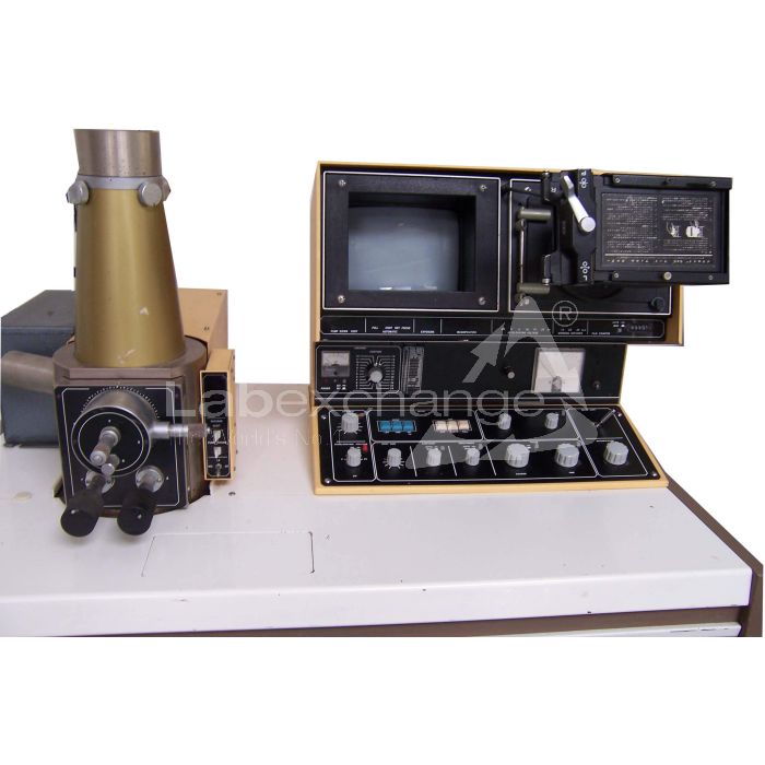
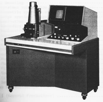
The Model T200 Scanning Microscope has been developed under the design philosophy of combining simple Operation, simple maintenance, and high performance. Accordingly, quality micrographs comparable to those obtainable by a large instrument can be readily obtained without any special skill.
These features together with the various advantages peculiar to the scanning microscope, such as very large depth of focus, wide magnification range, and minimal specimen preparation, make the T200 a most effective instrument for research work, quality control, and as a visual education aid.
GENERAL
The scanning electron microscope is a comparatively recent addition to the microscope family and is proving to be very popular along with the long established light and transmission type electron microscopes.
In spite of their merits and demerits, each type of microscope has its role to play depending on the field of application. For example, if a high resolving power and large depth of focus is not called for, a light microscope would be ideal. Where a very high resolving power is required, a transmission electron microscope would be necessary. However, when using a transmission electron microscope, the specimen must be very thin (less than 1 um (10,000 Ä) in thickness), a factor which requires a great deal of skill on the part of the user in order to prepare such specimens.
The scanning electron microscope, on the other hand, offers a fairly high resolution and moreover, since it is possible to use bulk specimens (specimen thickness being of no consequence), specimen preparation is easy. In addition to which, the depth of focus is large, thereby enabling 3-D observation.
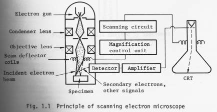
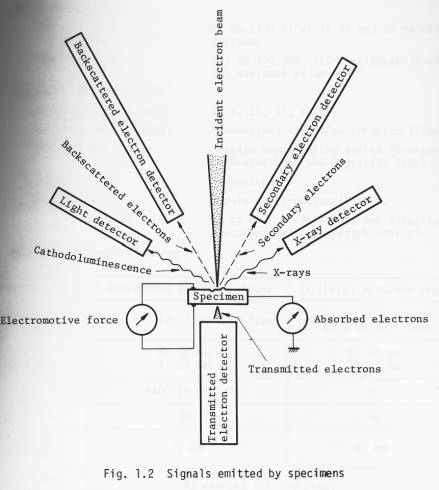
The operational principle of the scanning electron microscope is illustrated in Fig. 1.1. A finely focused electron probe is made to scan the specimen, resultant upon which, secondary and backscattered electrons, etc. (Fig. 1.2) are emitted from the surface of the specimen. These signals are then detected by a detector and outputted via an amplifier to a synchronously scanned CRT as an intensity variation signal. The CRT raster width divided by the electron probe scanning width gives the Image magnification.
By adding an appropriate detector (optional) to the Standard T200, transmitted electron images, cathodoluminescence images, etc. can be observed in addition to secondary and backscattered electron images. Further, by incorporating an X-ray detector, X-ray analysis becomes possible.
Specifications
Technical data
n Performance
Resolution: 10 nm (100 Ä) at 25 kV and 20 mm working distance.
Magnification: 15x to 100,000x (15x available at working distance 48 mm only).
n Electron optical system
Accelerating voltage: 2, 5, 10, 15, 25 kV.
Electron gun filament: Precentered cartridge tungsten filament.
Lens system: 3-stage demagnifying system (2-stage condenser lens and objective lens).
Alignment: Mechanical.
Stigmator: 8-pole electromagnetic type.
Image fine shift: Up to ±10 um (25 kV) in any direction; electromagnetic, joystick control.
n Specimen stage (twin stage)
|
Specimen stage |
I (Eucentric specimen stage) |
II (Large specimen stage) |
|
Specimen accomodation |
Up to 10 dia. x 10 thick*(mm) |
Up to 76.5dia. x 25.5 thick ** (mm) |
|
Range of movement |
X: 10 mm Y: 20 mm |
X: 40 mm Y: 40 mm |
|
Tilt |
-40° to +90°*** |
|
|
Rotation |
360° |
|
|
Working distance |
20 mm |
48 mm |
|
Specimen exchange |
By drawing out the stage |
|
|
Signal terminal |
Optional (max. 48 pins) |
|
* 32 dia., 51 dia., 76.5 dia. (mm) optionally available.
** Up to 127.5 dia. x 25.5 thick (mm) possible.
*** 220° possible.
n Scanning system
Secondary and backscattered
electron detection*: By a detector (comprizing a scintillator, a light pipe, a photomultiplier and a collector).
* Backscattered electron detector, which is capable of obtaining topographic and composition images, transmitted electron detector, cathodoluminescence detector, specimen current detector, X-ray
detector: Optionally available.
Scanning modes: Frame scan (including TV scan), line scan and Y modulation.
Scanning speeds: Visual TV scan; 0.2, 0.33, 10 sec/ frame.
Line scan 0.2, 0.33, 10 sec/frame.
Record 60 sec/frame.
Magnification: 35x to 100,000x (23 steps; series of 35,
50, 75, 100, 150, 200, 350, ).
(15x available at WD = 48 mm).
Viewing area: 135 mm x 180 mm.
Cathode ray tube: 230 mm, green phosphor CRT (used for viewing and recording*).
* A CRT exclusively used for recording is optionally available.
n TV signal output terminal : BNC-R connector; VTR available; composite video signal output; positive polarity; output voltage 1 Vp-p; use of 75 ohm coaxial cable; scanning
frequency horizontal: 15.75 kHz, vertical: 60 Hz.
n Recording System
(complete with electro-
magnetic shutter and
shutter button)
CSI-1: Standard; Brownie roll film; 1 to 1/2 photographing ratio.
CSI-2: Polaroid pack film; 1 to 3/4 photographing ratio (optional).
CSI-3: 35 mm roll film; 1 to 1/4 photographing ratio (optional).
CSI-4: Polaroid sheet film; 1 to l photographing ratio (optional).
n Vacuum system
Operation: Fully automatic.
Ultimate pressure: 7 x 10 -4 Pa (5 x 10 -6 Torr).
Vacuum gauge: Pirani gauge.
Pump-down time: About 30 minutes (from cold).
Specimen exchange: About 2.5 minutes.
Oil rotary pump: 100 l/min 1 unit.
Oil diffusion pump: 420 l/sec 1 unit.
n Safety devices: Devices for power failure, water failure and vacuum deterioration: built-in.
n Miscellaneous: Service outlet: built-in (100 V, 2 A; used for operating optional attachments).
The instrument can be wheeled an its own casters.
2. Installation requirements
n Power and water
Power: 100 V, 50/60 Hz, single phase, 2 kVA (basic instrument: 1.2 kVA; attachments: 0.8 kVA).
Starting current 60 A (0.2 sec.)
Fluctuation: less than ±10% (that also includes initial Operation).
Ground terminal: 1 terminal, less than 100 Ω.
Cooling water: Flow rate 2 l/min at 0.05 0.2 MPa (0.5 2 kg/cm 2 ).
Temperature 20 ±5 °C (water temperature at the outlet: not greater than 35 °C).
Faucet 1, 12 mm O.D.
Drain 1.
n Installation room
Room temperature: 20 ±5 °C.
Relative humidity: Less than 80%.
Floor vibration: Less than 2 µm p-p (5 Hz) in the X, Y and Z directions, less than 3 µm p-p (10 Hz) in the X, Y and Z directions, and less than 8 µm p-p (50 Hz) in the X, Y and Z directions (with the instrument installed).
Stray magnetic fields: Less than 0.3 µT (3 mG).
Note: The above specifications are subject to change without notice.
Layout, dimmensions and weight
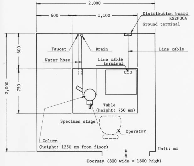
Overall weight (basic instrument: about 280 kg)
Votre Labexchange-équipe sera heureux de vous aider:

Christian Schmid
Laboratoire et analyse, Équipment de laboratoire, Life science

Hubert Sauter
Spectroscopie, Chromatographie


